If you’ve just been told you have a circumvallate placenta, the first question on your mind is almost certainly: will my baby be okay? We understand how frightening that moment can feel, and we want to give you a clear, honest answer right away. Many pregnancies with this diagnosis result in healthy, full-term deliveries – especially when the condition is detected early and managed by an experienced maternal-fetal medicine specialist. Close monitoring makes an enormous difference, and you are not alone in this.
Circumvallate placenta affects about 1-2% of pregnancies (the most-cited figure is around 1.2%). It has very few specific symptoms, which makes early detection challenging – but once identified, decisive action can protect both you and your baby.

What is a circumvallate placenta?
Circumvallate placenta (also called placenta circumvallate) involves the placenta – the organ responsible for delivering oxygen and nutrients to your baby while keeping your blood supplies separate.
Normally, the chorionic plate (the fetal side of the placenta) aligns with the basal plate (the maternal side). In a circumvallate placenta, the chorionic plate is too small, causing the fetal membranes to fold back on themselves around the placenta’s edge. This creates a raised ring or ridge that can constrict the umbilical cord and reduce the baby’s nutrient and blood supply.
The condition exists on a spectrum. In partial circumvallate placenta, only a portion of the placental edge is affected, and outcomes tend to be more favorable. In complete circumvallate placenta, the fold encircles the entire placental margin, which is associated with a higher risk of complications including antepartum hemorrhage and preterm birth. These folds can increase the risk of:
Placental abruption – premature separation of the placenta from the uterine wall
Fetal growth restriction (IUGR) and low birth weight
Oligohydramnios – insufficient amniotic fluid
Second trimester bleeding
Preterm birth or, in severe cases, miscarriage
Emergency C-section and neonatal intensive care unit (NICU) admission
The management of circumvallate placenta depends on the specific circumstances of each pregnancy, and a team approach – including high-risk obstetrics and antenatal surveillance – is often the key to a good outcome.
What causes circumvallate placenta?
No single definitive cause has been identified. The condition originates from abnormal placental implantation during the first trimester, and it can occur in women of any age, background, or lifestyle. Something we always stress: this is not your fault. In most cases, placental implantation issues come down to chance – not something you did or didn’t do.
Research does point to several associated factors that may raise the likelihood of abnormal implantation, including prior uterine surgery, multiparity (having had multiple pregnancies), and the presence of a subchorionic hematoma in early pregnancy. Prior uterine procedures – such as a D&C, myomectomy, or cesarean section – may alter the uterine lining in ways that affect how the placenta anchors itself. Multiparity may play a role because repeated pregnancies can change the uterine environment over time. A subchorionic hematoma, which is a small bleed between the placenta and the uterine wall, may interfere with normal placental folding and membrane attachment in those critical early weeks. A retrospective study published in PMC also notes that circumvallate placenta is more commonly identified in pregnancies already under close surveillance for other reasons – which is why comprehensive prenatal care matters so much.
Because circumvallate placenta is often asymptomatic in its early stages, many cases are only discovered during a detailed ultrasound exam. This is one reason why state-of-the-art imaging and regular monitoring are so valuable – catching the condition early gives your care team the best possible opportunity to track placental function and fetal growth closely throughout your pregnancy.
Can we prevent a circumvallate placenta?
Because there is no definitive cause, we can’t guarantee prevention. The risk factors overlap with those for other placental complications, such as placenta previa or pre-eclampsia. What we can do is follow general guidelines that support a healthy pregnancy and allow early detection:
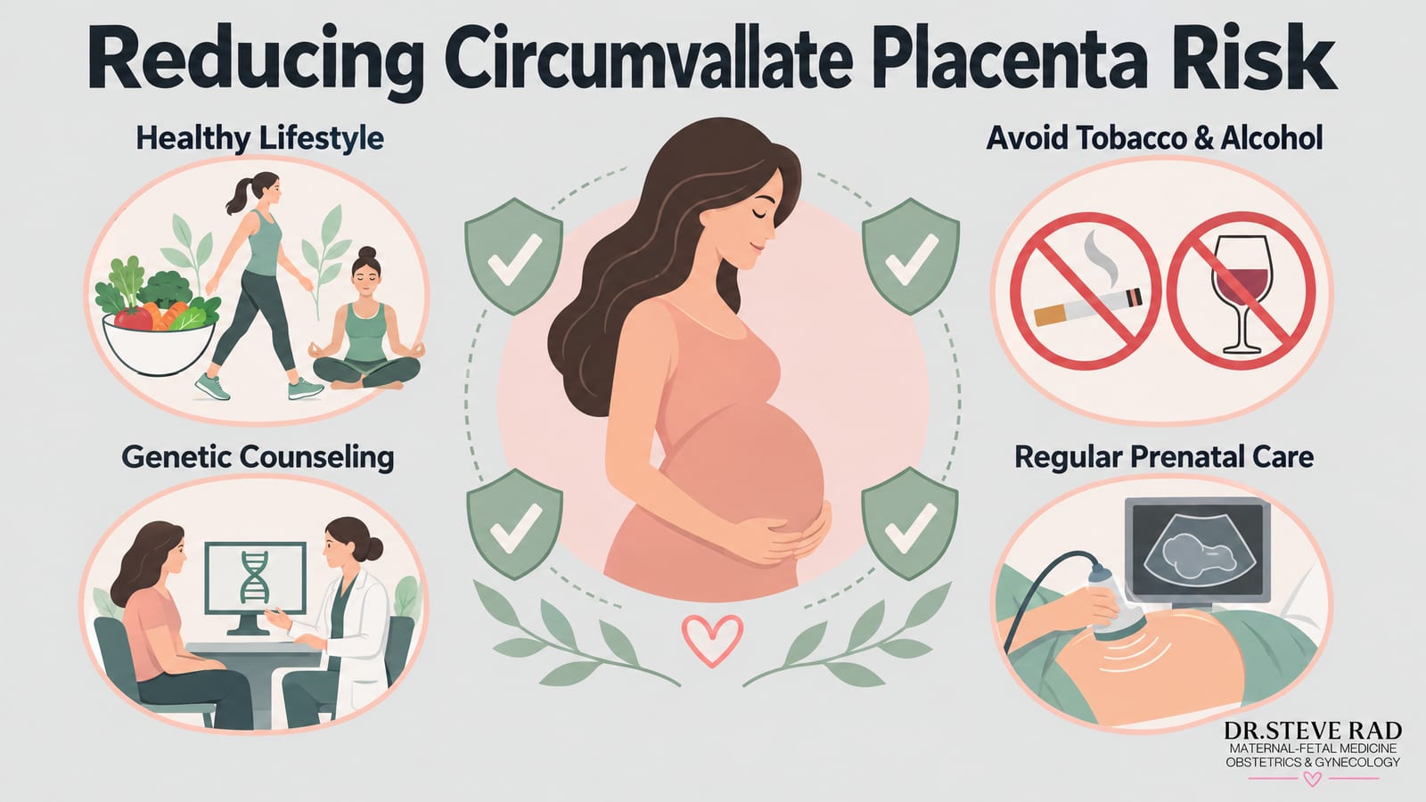
A balanced diet, appropriate physical activity, and stress management before and during pregnancy
Avoiding tobacco and excessive alcohol
Genetic counseling for women with a family history of placental abnormalities
Regular prenatal care – check-ups, ultrasounds, and prenatal testing – to detect circumvallate placenta early and keep its impact contained
Even with the best prenatal care, some conditions still occur. But early detection and expert management can make a profound difference in how the pregnancy develops.
How does circumvallate placenta affect pregnancy?
A circumvallate placenta raises the incidence of several serious perinatal complications – but it does not make any of them inevitable. The distinction between partial and complete circumvallate placenta matters enormously here: partial cases, where only a portion of the placental edge is folded back, tend to carry a milder risk profile and are more likely to result in a healthy, full-term delivery. Complete circumvallate placenta, where the fold encircles the entire placental margin, is associated with a higher likelihood of complications and warrants closer surveillance throughout pregnancy. The degree of risk also depends on how early the condition is detected and the quality of antenatal monitoring.

Call (844) 473-6100 or click here to schedule online
Placental insufficiency and fetal growth restriction
When the folded placental edge restricts blood flow, the fetus receives less oxygen and fewer nutrients – a condition known as intrauterine growth restriction (IUGR). Babies affected by IUGR face an elevated risk of low birth weight and longer NICU stays, and in the most severe cases, outcomes can be serious. That said, many babies diagnosed with IUGR in the context of circumvallate placenta go on to do well, particularly when the condition is caught early and monitored closely – and that close monitoring is exactly what Dr. Rad’s concierge approach is built around.
During growth scans, Dr. Rad looks specifically for slowing fetal measurements, reduced amniotic fluid, and abnormal Doppler flow in the umbilical artery. These are the early warning signs that matter most – changes that, when identified in time, open a window for intervention before the situation becomes critical. Serial Doppler ultrasound and biophysical profile assessments allow us to build a detailed picture of how your baby is growing week by week, so nothing is missed.
If you have already been seen in the ‘How does circumvallate placenta affect pregnancy?’ section of this page, the core mechanism is the same: a constricted placental margin limits the surface area available for nutrient exchange. What matters here is what we do about it. State-of-the-art 3D and 4D ultrasound technology, available through Dr. Rad’s practice, gives us a level of detail that standard imaging simply cannot match – and 24/7 availability means that if something changes between scheduled appointments, you are never left waiting for answers. That is the Dr. Rad difference.
Placental abruption
Placental abruption – where the placenta separates from the uterine wall before delivery – is a serious emergency that can lead to maternal hemorrhage, preterm delivery, or, in the most severe cases, stillbirth. With a circumvallate placenta, the folded membrane edge creates mechanical tension that elevates this risk. The degree of risk depends largely on how complete the circumvallate folding is and how closely the pregnancy is monitored; partial cases managed with regular surveillance often progress far better than the diagnosis initially suggests.
That reassurance is more than theoretical. One of Dr. Rad’s patients, Shanley, experienced placental abruption during her pregnancy – one of the most frightening complications a mother can face – and went on to deliver a healthy baby. Her story is a powerful reminder that expert, timely intervention genuinely changes outcomes. As Dr. Rad often says, knowing the warning signs, acting quickly, and having a specialist in your corner make all the difference. That is the Dr. Rad difference.
If you have been diagnosed with circumvallate placenta and are worried about abruption risk, the most important step you can take is ensuring you have a maternal-fetal medicine specialist monitoring your pregnancy closely. Dr. Rad provides 24/7 availability – including home visits – so that if you experience sudden abdominal pain, heavy bleeding, or reduced fetal movement, you are never left waiting for answers during a moment that cannot wait.
Preterm premature rupture of membranes (PPROM)
Circumvallate placenta makes the amniotic membranes smaller and more fragile, heightening the risk of PPROM – rupture before 37 weeks. The consequences depend heavily on gestational age at delivery. PPROM in the second trimester carries the greatest risk, including severe antepartum hemorrhage and chorioamnionitis (infection of the placental tissues). PPROM closer to term is far more manageable and often results in a slightly early but otherwise healthy delivery. Neonatal survival rates for PPROM near term are reassuringly high – outcomes improve significantly with each additional week of gestation gained, which is why the goal of close monitoring is always to keep the pregnancy progressing as long as it is safely possible to do so. Umbilical cord compression is an additional concern when amniotic fluid levels drop.
Circumvallate placenta survival rate and outcomes
If you’ve just received this diagnosis, the first thing we want you to know is this: many pregnancies with a circumvallate placenta result in healthy babies. That’s not false reassurance – it’s what the research consistently shows, and it’s important context for everything that follows.
Circumvallate placenta affects roughly 1-2% of pregnancies. In those cases, outcomes vary widely depending on how much placental edge is involved and how early the condition is identified. When only a partial fold is present, many women carry to full term without significant complications. A complete circumvallate placenta carries higher risks, but even then, neonatal survival rates are generally favorable when the pregnancy is closely monitored by a maternal-fetal medicine specialist.
Here’s an honest look at the elevated risks associated with this condition:
Preterm birth: Studies suggest preterm delivery occurs in roughly 30-50% of symptomatic cases, though many preterm babies born after 34 weeks do very well with appropriate neonatal support.
Intrauterine growth restriction (IUGR): Reduced placental surface area can limit fetal growth, which is why regular growth ultrasounds are so critical for tracking fetal well-being.
Placental abruption: The risk is higher than in typical pregnancies, but careful monitoring allows timely intervention.
Perinatal mortality: In severe, unmonitored cases, perinatal mortality rates have been reported as higher than average – but with specialist oversight, this risk is substantially reduced.
The takeaway? A circumvallate placenta diagnosis is serious, but it is not a worst-case scenario. With the right care team, close surveillance, and a clear management plan, the odds of bringing home a healthy baby are genuinely encouraging. That kind of personalized, proactive care makes all the difference.
Circumvallate placenta survival rate and outcomes
This is the question we hear most often, and you deserve a direct answer. The published data are genuinely reassuring for most patients.
Studies show that the majority of pregnancies complicated by circumvallate placenta result in a live birth, particularly when the condition is detected and managed under specialist care. The elevated risks are real but quantifiable:
Preterm birth is the most common adverse outcome, occurring at a significantly higher rate than in uncomplicated pregnancies – but most preterm infants born after 28 weeks, and virtually all born after 32 weeks, survive with appropriate NICU support.
Perinatal mortality (stillbirth and early neonatal death) is elevated compared to the general population, but remains a minority outcome. The absolute risk is highest in cases of complete circumvallate placenta complicated by severe IUGR or early placental abruption.
NICU admission is more common than in uncomplicated pregnancies, primarily due to prematurity and low birth weight – but a NICU stay, while stressful, is not the same as a poor long-term outcome. Most of these babies go home healthy.
Full-term delivery is entirely possible, especially with partial circumvallate placenta and no secondary complications. Many patients with this diagnosis deliver vaginally at or near term.
Can the condition resolve or stabilize? The placental anatomy itself does not change after implantation, but you can actively manage the risks it creates. Growth scans and Doppler ultrasound track whether fetal growth is staying on a healthy curve, whether amniotic fluid is adequate, and whether placental blood flow is deteriorating. Biophysical profile assessments add another layer of reassurance between appointments. The goal of antenatal surveillance is to catch any deterioration early – long before it becomes an emergency.
The honest prognosis: with close monitoring by a maternal-fetal medicine specialist, most women with a circumvallate placenta bring home a healthy baby. The diagnosis calls for vigilance, not despair.
How is circumvallate placenta treated?
Treatment is tailored to each pregnancy. Many cases are managed through expectant management – close monitoring without immediate intervention – combined with lifestyle adjustments such as modified activity or a carefully monitored diet to support fetal growth.
Frequent antenatal surveillance (growth scans, Doppler ultrasound, biophysical profiles, and amniotic fluid assessments) allows our team to track the baby’s well-being and respond quickly if anything changes. If early delivery becomes necessary, gestational age is the critical variable. Corticosteroids are typically administered between 24 and 34 weeks of gestation to accelerate fetal lung maturity and meaningfully improve neonatal survival. Tocolytic medications may be used short-term to delay delivery and allow corticosteroids time to work.
If complications escalate, labor induction or an emergency C-section may be the safest path. High-definition imaging has been central to some of our most successful outcomes – including Angelica, a patient from the Philippines who was diagnosed with circumvallate placenta while visiting the U.S. After seeking Dr. Rad’s care, she became the proud mother of a healthy baby girl.
When to seek specialist care for circumvallate placenta
Receiving a circumvallate placenta diagnosis can feel overwhelming, but knowing exactly when to reach out for help puts you back in the driver’s seat. While many pregnancies with this condition are managed successfully with routine monitoring, there are certain warning signs that mean you should contact your care team right away – don’t wait for your next scheduled appointment.
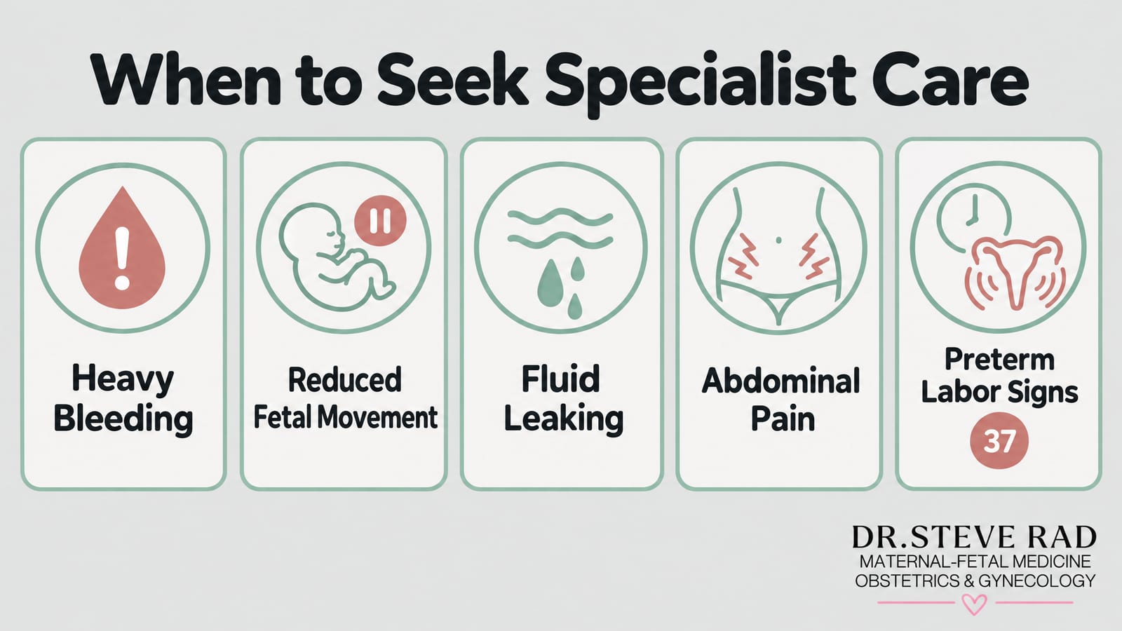
Reach out immediately if you experience any of the following:
Heavy or bright red vaginal bleeding – any significant bleeding during pregnancy warrants urgent evaluation, as it can signal placental abruption, a known risk with circumvallate placenta.
Reduced or absent fetal movement – if your baby is moving less than usual or you haven’t felt movement in several hours, call your provider straight away.
Sudden gush or steady trickle of fluid – this could indicate premature rupture of membranes (PPROM), which carries serious risks at earlier gestational ages.
Persistent abdominal pain or cramping – especially if it feels different from normal pregnancy discomfort or comes with back pain.
Signs of preterm labor – regular contractions before 37 weeks should always be assessed promptly.
If your current provider isn’t offering the level of monitoring you feel you need, it may be worth exploring a high-risk pregnancy second opinion. As a maternal-fetal medicine specialist, Dr. Rad provides 24/7 access – because when something feels wrong, you deserve answers immediately, not at the next available slot. That is the Dr. Rad difference.
Why trust Dr. Rad with your pregnancy?
Even a pregnancy that begins as low-risk can become a high-risk obstetrics case. Double-board-certified Dr. Steve Rad is an obstetrician-gynecologist with subspecialty training in perinatal maternal-fetal medicine, and has been named a top maternal-fetal medicine specialist in Los Angeles by Los Angeles Magazine for six consecutive years.
We treat all patients as VIPs, ensuring you feel cared for and supported at every step. We understand this diagnosis can feel terrifying – our role is to give you the information and the care you need to feel supported every step of the way. That is the Dr. Rad difference.
Our offices are located throughout Southern California – Beverly Hills, Santa Monica, West Los Angeles, West Hollywood, Culver City, Hollywood, Venice, Marina del Rey, Malibu, Manhattan Beach, Newport Beach, Irvine, and downtown Los Angeles. We also offer in-home prenatal care, a fly-in program for out-of-town and international patients, and virtual consultations worldwide.
Book your appointment online or call us anytime at (844) 473-6100.
Call (844) 473-6100 or click here to schedule online
Sources
HD live imaging of circumvallate placenta – AboEllail – 2015 – Ultrasound in Obstetrics & Gynecology – Wiley Online Library
https://doi.org/10.1002/uog.14839
Circumvallate Placenta: Associated Clinical Manifestations and Complications-A Retrospective Study
https://www.ncbi.nlm.nih.gov/pmc/articles/PMC4248327/
Premature Rupture of Membranes
https://www.ncbi.nlm.nih.gov/books/NBK532888/
Restriction (IUGR): etiology and diagnosis. Curr Obstet Gynecol Rep 2013; 2:102-111. Dukatz R, Henrich W, Entezami M, Nasser S, Siedentopf
https://www.degruyter.com/document/doi/10.1515/crpm-2020-0020/html