Cervical incompetence (also known as cervical insufficiency) affects about 1% of all pregnancies and accounts for 8% of second-trimester losses in high-risk women. Because the condition is often painless, many patients only learn of it through a routine ultrasound – which is exactly why understanding what those sonographic findings mean is so important.
Dr. Steve Rad is a double-board-certified Obstetrician-Gynecologist with subspecialty training in Maternal-Fetal Medicine (MFM). His team at Los Angeles Fetal and Maternal Care has decades of perinatology experience and provides state-of-the-art diagnosis and treatment for cervical insufficiency. We understand pregnancy can be a vulnerable and anxious time – and we are here to guide you through every step.

What is cervical incompetence?
Cervical incompetence is the structural inability of the cervix to remain closed and support a growing fetus. The cervix begins to dilate and efface (shorten and thin) during the second trimester before the pregnancy reaches term, which can lead to a spontaneous abortion around the fourth or fifth month. According to the American College of Obstetricians and Gynecologists (ACOG), this affects roughly one pregnancy in every 100.
Cervical incompetence risk factors
Though the cause of this condition is often unknown, several factors are known to raise a woman’s risk of cervical insufficiency:
Second-trimester pregnancy loss in a prior pregnancy
Connective tissue disorders such as Ehlers-Danlos syndrome or Marfan’s syndrome
In utero exposure to diethylstilbestrol (DES)
History of cervical procedures, including cone biopsy or LEEP
Cervical trauma from a prior delivery, D&C, or other procedure
Second-trimester pregnancy loss
Women who have miscarried in the second trimester of a previous pregnancy are at twice the risk for cervical insufficiency in future pregnancies. This history is one of the strongest clinical indicators Dr. Rad uses when deciding how closely to monitor cervical length via transvaginal ultrasound in a subsequent pregnancy.
Connective tissue disorders
Genetic conditions that affect collagen – such as Ehlers-Danlos syndrome and Marfan’s syndrome – weaken cervical tissue and increase insufficiency risk. Because the cervix relies on collagen for its structural integrity, patients with these diagnoses benefit from early and regular cervical length surveillance throughout the second trimester.
Exposure to diethylstilbestrol (DES)
DES is a synthetic estrogen once prescribed during pregnancy. In utero DES exposure has been directly linked to cervical insufficiency in daughters born to women who took it. DES-exposed patients often have a structurally abnormal cervix that may appear shorter or differently shaped on ultrasound, making imaging surveillance especially important for this group.
History of cervical procedures
Women who have had precancerous cervical lesions removed via cone biopsy or LEEP may have reduced cervical length and structural integrity, raising their risk of insufficiency. The amount of tissue removed during these procedures directly influences how much functional cervical length remains – a detail Dr. Rad carefully factors into your personalized monitoring plan.
Cervical trauma
Tears from a prior delivery, dilation and curettage (D&C), or other cervical procedures can compromise the cervix’s ability to stay closed during a subsequent pregnancy. Even when trauma appears minor at the time, scar tissue can alter the cervix’s mechanical strength in ways that only become apparent on a detailed transvaginal ultrasound assessment.

Race
Black women are more prone to cervical incompetence.
Uterine abnormalities
Structural uterine abnormalities are a well-established risk factor for cervical incompetence and are often identified during a comprehensive sonographic evaluation. Conditions such as a bicornuate or septate uterus can place uneven mechanical pressure on the cervix, increasing the likelihood of early dilation. State-of-the-art 3D ultrasound imaging allows Dr. Rad to assess both uterine structure and cervical length in a single, detailed examination – that is the Dr. Rad difference.
Signs of cervical incompetence
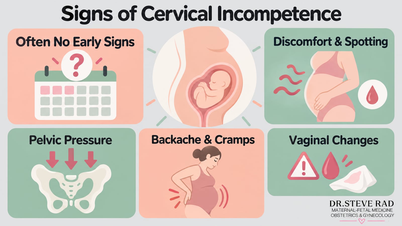
Symptoms do not always appear in early pregnancy, which is why regular prenatal monitoring with Dr. Rad is so important. When signs do occur, they may include:
Mild discomfort or spotting between 14 and 20 weeks of gestation
A sense of pelvic pressure
Backache
Abdominal cramps
Vaginal discharge
Light vaginal bleeding
How to diagnose cervical incompetence
Cervical insufficiency cannot be detected outside of pregnancy, and even during pregnancy it can be difficult to identify – particularly in a first pregnancy. Dr. Rad will review your symptoms and full obstetric history, paying close attention to any prior second-trimester losses or painless cervical dilation.
Clinical criteria that support a diagnosis include:
A history of painless cervical dilation and second-trimester delivery
Advanced cervical dilation and effacement before 24 weeks, without infection, contractions, bleeding, or rupture of membranes
Prior cervical laceration during labor and delivery
The primary diagnostic tool is transvaginal ultrasound, which lets Dr. Rad measure cervical length and identify specific sonographic changes at the internal os. Screening typically occurs between 16 and 24 weeks of gestational age, when cervical shortening is most clinically meaningful.
During a transvaginal ultrasound evaluation, Dr. Rad looks for several key findings that together paint a clear picture of cervical health. The most important measurement is cervical length – a cervix measuring 25 mm or less is considered short and raises concern for insufficiency, while a length below 20 mm warrants close monitoring and often intervention. A normal cervical length at mid-pregnancy is typically 35-48 mm, so even a modest shortening can carry real clinical significance.
Beyond raw length, Dr. Rad also evaluates the shape and behavior of the internal os – the opening at the top of the cervix closest to the uterus. In a healthy pregnancy, this remains firmly closed, but with cervical insufficiency it can begin to open prematurely, creating what is called funneling. Funneling describes the characteristic V- or U-shaped widening of the internal os, where amniotic membranes begin to bulge downward into the cervical canal. In more advanced cases, the membranes may visibly balloon into the vagina – a finding that signals an urgent situation requiring immediate attention. These sonographic changes can appear before any symptoms develop, which is exactly why ultrasound screening is so valuable for patients with a history of pregnancy loss or known risk factors.
If your transvaginal ultrasound reveals a short cervix or early funneling, Dr. Rad will explain what those findings mean for your situation and outline a personalized care plan right away. Depending on gestational age and the degree of change observed, next steps may include increased monitoring, progesterone therapy to support cervical integrity, or a cerclage procedure to physically reinforce the cervix. You will never be left wondering what your results mean – that personal touch and clear communication is at the heart of the Dr. Rad difference.

Call (844) 473-6100 or click here to schedule online
Testing for cervical incompetence
Two key tests are used during the second trimester to evaluate cervical incompetence: transvaginal ultrasound and pelvic exam.
Transvaginal ultrasound
A transvaginal ultrasound is the gold standard for assessing cervical length and identifying early signs of insufficiency. The probe is gently placed in the vagina to obtain a clear, close-up view of the cervix and internal os – far more precise than a transabdominal scan.
A normal cervical length at mid-pregnancy is approximately 35-48 mm. A measurement of 25 mm or less is the key threshold: at this length, the risk of preterm birth rises significantly, and intervention is typically considered. Lengths below 20 mm carry an even higher risk and often prompt immediate action.
Beyond length, Dr. Rad evaluates the shape and behavior of the internal os. In a healthy cervix, the internal os remains closed, and the cervical canal is uniform. In cervical incompetence, the internal os begins to open, and the membranes can herniate upward into the canal, creating a characteristic funnel shape. This funneling is described as U-shaped (a wide, rounded opening) or V-shaped (a narrower, pointed opening) – both are abnormal sonographic findings. As the condition progresses, the membranes may balloon through the os, a sign that delivery may be imminent without intervention.
Understanding what these findings mean for your care plan is just as important as the measurements themselves. If Dr. Rad identifies a cervical length approaching 25 mm, he may recommend progesterone supplementation – a well-established first-line option that helps reduce the risk of preterm birth in women with a short cervix. When funneling is present alongside significant shortening, or if the cervix has dropped below 20 mm, a cerclage procedure may be the next step. The specific findings on your ultrasound – the degree of shortening, the shape of the funnel, and whether any ballooning is visible – directly inform which intervention is most appropriate for you and at what point in your pregnancy it should happen.
Because cervical shortening can progress rapidly, serial cervical length monitoring – repeat transvaginal ultrasounds every one to two weeks – is often recommended for women at elevated risk. This allows Dr. Rad’s team to track changes over time and act before the cervix shortens to a critical threshold.
We understand that receiving an abnormal cervical length result can feel frightening. At the Maternal Fetal Care Center, we walk you through every measurement and every finding in plain language, so you always know exactly where things stand and what comes next. That is the Dr. Rad difference – state-of-the-art diagnostics paired with the personal touch of a physician who is available to you 24/7.
What ultrasound findings indicate cervical incompetence?
When your provider orders a cervical incompetence ultrasound, they are looking for a handful of very specific sonographic markers that signal your cervix may not be holding up under the pressure of a growing pregnancy. Understanding what these findings mean can help you feel less anxious when you hear terms like “short cervix” or “funneling” during your appointment.
The most important measurement is cervical length. A healthy cervix typically measures 35-48 mm during the second trimester. A length at or below 25 mm is considered short and raises concern for cervical insufficiency. The shorter the measurement, the higher the risk of preterm birth – a cervix measuring under 15 mm is treated as a significant warning sign that usually prompts immediate action.
Beyond length, your specialist will look for funneling – a change in the shape of the internal os (the upper opening of the cervix). Normally, the internal os stays closed and looks like a straight line on ultrasound. With cervical incompetence, it can open into a V-shape or widen further into a U-shape, meaning the upper portion of the cervix has already begun to dilate. A related finding called beaking describes the very early stage of this process, where the internal os just begins to wedge open.
In more advanced cases, membrane prolapse or ballooning may be visible, where the amniotic sac begins to bulge through the dilating cervix. This serious finding often calls for emergent intervention. If you are navigating a high-risk pregnancy, having these findings evaluated by a maternal-fetal medicine specialist using state-of-the-art ultrasound technology ensures nothing is missed – that is the Dr. Rad difference.
Pelvic exam
A pelvic exam can reveal whether the amniotic sac is protruding through the cervical opening (prolapsed fetal membranes), a serious finding that often signals imminent preterm delivery. While transvaginal ultrasound remains the primary diagnostic tool, a pelvic exam provides an important complementary picture – particularly when symptoms such as pelvic pressure or unusual discharge suggest that changes to the cervix may already be advanced.
What ultrasound findings indicate cervical incompetence?
When Dr. Rad reviews your transvaginal ultrasound, he is looking for a specific set of sonographic markers that together paint a picture of cervical competence – or the lack of it:
Short cervical length: A cervix measuring 25 mm or less between 16 and 24 weeks is the primary quantitative threshold for concern. Below 20 mm, the risk of previable delivery before 24 weeks rises sharply.
Funneling at the internal os: The internal os opens, and the amniotic membranes herniate into the cervical canal, forming a U-shaped or V-shaped funnel. The funnel length and width help gauge severity.
Membrane prolapse and ballooning: In advanced cases, membranes bulge visibly through the external os – a finding associated with a high risk of PPROM (preterm premature rupture of membranes) and infection such as chorioamnionitis.
Cervical effacement: Progressive thinning and shortening of the cervical canal on serial scans, even without overt funneling, can indicate evolving insufficiency.
Understanding these findings helps you and your care team make informed, timely decisions. A short cervix on ultrasound is not a verdict – it is a signal that prompts a proactive care plan.

Imaging tests
If the fetal membranes are visible, an ultrasound may show signs of inflammation.
Cervical insufficiency complications
Left untreated, cervical incompetence identified on ultrasound can have serious consequences. The most significant risk is previable delivery – birth before 24 weeks of gestational age, when survival outside the womb is extremely unlikely. At this stage of pregnancy, a baby’s lungs, brain, and other vital organs are simply not developed enough to sustain life, making early detection through transvaginal ultrasound genuinely life-saving.
Other complications include preterm premature rupture of membranes (PPROM), intrauterine infection (chorioamnionitis), and significant neonatal morbidity in babies born very early. Infants delivered prematurely due to cervical insufficiency face elevated risks of respiratory distress syndrome, intraventricular hemorrhage, long-term developmental delays, and extended stays in the neonatal intensive care unit – challenges that can affect the entire family for years to come.
It is also worth understanding that cervical insufficiency rarely causes the kind of painful contractions associated with typical preterm labor. Many patients feel little to nothing as the cervix silently shortens and dilates – which is precisely why routine ultrasound monitoring is so important for anyone with known risk factors.
By the time symptoms like pelvic pressure or spotting appear, the situation may already be advanced. This is why prompt action after an abnormal ultrasound finding is so critical, and why Dr. Rad’s team prioritizes close, personalized surveillance for every high-risk pregnancy under their care. That is the Dr. Rad difference.
What is the cervical cerclage procedure?
Cervical cerclage – sometimes called a cervical stitch – is the primary surgical treatment for cervical insufficiency. A suture is placed around the cervix to keep it closed, then removed at approximately 37 weeks. Cerclage candidacy is directly tied to what your cervical incompetence ultrasound reveals: women with a cervical length at or below 25 mm, or with sonographic evidence of funneling or internal os changes, are typically evaluated for the procedure. In other words, your ultrasound findings are not just a diagnostic tool – they actively shape your treatment plan.
The cervical cerclage procedure is effective 85-90% of the time.

The optimal timing is at the end of the first trimester (12-14 weeks). When cerclage is needed later in pregnancy due to acute cervical changes, it is called an emergent cerclage. Women who require emergent cerclage will typically need a cerclage placed prophylactically in subsequent pregnancies.
Transabdominal cerclage is reserved for patients who cannot have a transvaginal cerclage – for example, those with a very short or surgically altered cervix. In this approach, the surgeon places the suture through the abdomen. It carries additional risks, including rupture of membranes, chorioamnionitis, and intraoperative hemorrhage from the uterine veins.
For women whose ultrasound shows a shortened cervix but who are not yet cerclage candidates, progesterone therapy is an important first-line option. Vaginal progesterone has been shown to reduce the risk of preterm birth in women with a short cervix identified on ultrasound, and Dr. Rad will discuss whether this approach – alone or alongside cerclage – is right for your specific situation. For women who are not cerclage candidates, complete bed rest remains an alternative management strategy.
Progesterone therapy for cervical insufficiency
Alongside cerclage, progesterone therapy is an important treatment option for women with a short cervix identified on ultrasound. Vaginal progesterone is commonly prescribed for women with a cervical length of 25 mm or less who have not had a prior preterm birth, as it has been shown to reduce the risk of preterm birth in this group. For women with a history of spontaneous preterm birth, weekly progesterone injections (17-OHPC) may be recommended beginning in the second trimester. Dr. Rad will review your ultrasound findings and full history to determine whether progesterone therapy, cerclage, or a combination of both is the right approach for your care plan.
Cervical incompetence prevention
Cervical incompetence cannot be prevented, but healthy habits and consistent prenatal care give you the best chance of a full-term pregnancy:
Attend frequent prenatal visits and report any new symptoms promptly – early detection through serial cervical length monitoring can make all the difference.
Maintain good nutrition, including folic acid, calcium, and iron. A daily prenatal vitamin started before conception helps fill nutritional gaps.
Aim for healthy pregnancy weight gain – typically 25 to 35 pounds for women of normal pre-pregnancy weight.
Avoid smoking, alcohol, and illicit drugs, and check with Dr. Rad before taking any supplements.
Call (844) 473-6100 or click here to schedule online
Cervical incompetence FAQs
What does cervical insufficiency feel like?
Cervical insufficiency often does not cause noticeable physical sensations or discomfort. Instead, cervical insufficiency is usually diagnosed based on certain signs and symptoms that may include:
- Painless Cervical Dilation
- Vaginal Pressure
- Increased Vaginal Discharge
- Backache or Pelvic Pressure
What causes cervical insufficiency?
Cervical insufficiency occurs when the cervix begins to dilate or efface (thin out) prematurely. The exact cause is not always clear, but several factors and potential contributors have been identified:
- Congenital Weakness
- Previous Cervical Trauma or Surgery
- Uterine Abnormalities
- Multiple Pregnancies
- Hormonal Factors
- Infections
- Previous Preterm Birth
How common is cervical insufficiency?
It’s estimated that cervical insufficiency occurs in about 1% to 2% of all pregnancies. However, the exact prevalence may vary depending on the population studied and the diagnostic criteria used.
Will frequent ultrasounds prevent cervical insufficiency?
Frequent ultrasounds may be used as part of the monitoring and assessment of cervical insufficiency but are not considered a preventive measure on their own. Ultrasound is often used in conjunction with clinical evaluation, cervical cerclage, and medications.
Can a doctor tell if you have cervical insufficiency?
Yes, a doctor can often diagnose cervical insufficiency, also known as an incompetent cervix, through a combination of clinical evaluation, medical history, and various diagnostic tests such as:
- Medical History
- Physical Examination
- Cervical Length Assessment
- Cervical Ultrasound
- Previous Pregnancy History
- Cervical Biopsy
Can we reverse cervical insufficiency?
While cervical insufficiency cannot be completely reversed, there are medical interventions and treatments that can help manage and reduce the risk of complications. Though, the effectiveness of interventions may vary from person to person.
Does misoprostol cause cervical insufficiency?
There is no direct evidence to suggest that misoprostol causes cervical insufficiency.
How can you prevent cervical insufficiency?
Cervical insufficiency cannot always be prevented as it is often related to structural or anatomical factors. However, there are some strategies that may help reduce the risk of cervical insufficiency or identify it early for appropriate management:
- Preconception Counseling
- Cervical Length Assessment
- Cervical Cerclage
- Progesterone Supplementation
- Lifestyle Factors
- Prenatal Care
Is cervical insufficiency hereditary?
Cervical insufficiency is not typically considered a strongly hereditary condition. It is a complex condition, and its exact causes can vary from person to person.