Receiving a diagnosis involving your placenta can feel overwhelming. The good news is that most placenta problems, including the rarest ones, can be managed effectively when the right specialist identifies them early. This guide covers both common and rare placenta complications, including vasa previa, circumvallate placenta, placenta accreta spectrum disorders, and placental insufficiency, so you understand what each condition means and when to seek expert care.
Almost 4% of all pregnancy complications involve a placental condition. With rising C-section rates and increasing use of IVF, the incidence of several rare placenta problems, particularly placenta accreta spectrum disorders, is climbing. That makes specialist awareness more important than ever.
Dr. Steve Rad is a double-board-certified Obstetrician-Gynecologist with subspecialty training in Maternal-Fetal Medicine (MFM). As a perinatologist at the Los Angeles Fetal and Maternal Care Center, he and his team have decades of experience guiding patients through exactly these diagnoses, with compassion and clinical precision. That is the Dr. Rad difference.
What is the placenta?
The placenta is an organ that develops in the uterus during pregnancy. It delivers oxygen and nutrients to your baby and removes waste from your baby’s blood through the umbilical cord. When the placenta develops abnormally, attaches incorrectly, or separates too early, the consequences for mother and baby can be serious.
Placenta previa
Placenta previa occurs when the placenta attaches lower than normal in the uterus, partially or completely covering the cervix. It affects 1 in 200 pregnancies and usually becomes apparent in the third trimester. A low-lying placenta detected earlier in pregnancy may resolve through placental migration as the uterus grows, but complete previa rarely does.
Management depends on severity. Asymptomatic cases are often handled with expectant management, including bed rest and pelvic rest, to reduce bleeding risk. This conservative approach allows the pregnancy to continue as long as possible under close monitoring, with serial ultrasounds used to track placental position and fetal well-being. However, when bleeding becomes heavy or uncontrolled, the situation shifts quickly – emergency intervention, including urgent Cesarean delivery, may be necessary to protect both mother and baby. Any vaginal bleeding requires prompt evaluation. Because vaginal delivery is unsafe when the placenta covers the cervix, most patients deliver by Cesarean section. Severe hemorrhage may require a blood transfusion.
Types of placenta previa
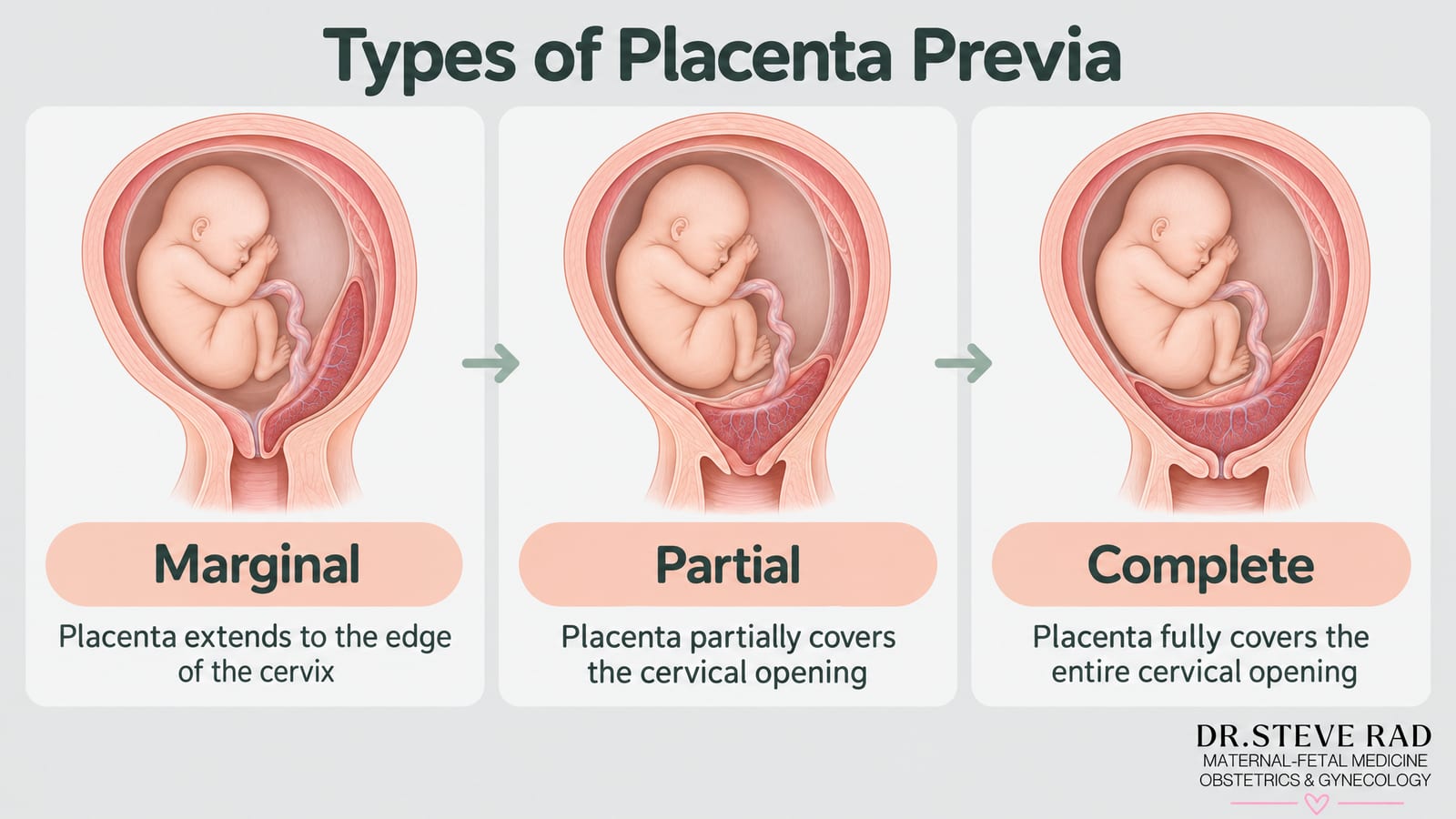
Marginal – The placenta extends to the edge of the cervix
Partial – The placenta partially covers the cervix
Complete – The placenta fully covers the cervix
Placenta previa risk factors
Over 35 years of age
Smoking
Prior C-section or uterine surgery
Carrying multiple fetuses (twins, triplets, etc.)

Placental abruption
Placental abruption is a medical emergency in which the placenta separates from the uterine wall before delivery. It occurs in about 1% of pregnancies and is a leading cause of premature delivery, newborn mortality, and fetal growth restriction (FGR). In severe cases, it can lead to stillbirth. When abruption occurs, time is everything – the placenta is your baby’s lifeline, and even a partial separation can rapidly compromise oxygen and nutrient delivery. Mothers may experience vaginal bleeding, abdominal pain, continuous contractions, and fetal heart abnormalities. It is worth noting that abnormal fetal heart rate patterns are a consequence of abruption rather than a cause – they signal that the baby is already under stress and that urgent specialist intervention is needed. Dr. Rad’s team has guided patients like Shanley through this frightening diagnosis, providing the close monitoring and rapid intervention that makes the difference in outcomes. Shanley’s experience is a powerful reminder of the Dr. Rad difference: concierge-level, 24/7 access to a renowned perinatologist who treats every patient as a VIP, not just a chart number.
Placental abruption risk factors
The following are true risk factors – conditions or behaviors that increase the likelihood of abruption occurring in the first place. This list does not include complications that arise from abruption, such as abnormal fetal heart rate patterns.
Abdominal trauma
Preterm membrane rupture
Smoking by either parent
Use of cocaine or alcohol
Prior placental abruption
Placenta previa
Uterine defects
High blood pressure or pre-eclampsia
Multiple fetuses
Previous Cesarean section
It is also important to understand that certain rare placenta problems – such as circumvallate placenta – are sometimes listed alongside abruption in clinical discussions, but they are better understood as separate conditions that can share overlapping symptoms like bleeding and preterm labor. If you have been told you have any unusual placental finding, that is exactly the kind of situation where a maternal-fetal medicine specialist like Dr. Rad can provide the expert, personalized guidance you deserve.

Call (844) 473-6100 or click here to schedule online
What is placental insufficiency?
Occurring in approximately 1 in 300 pregnancies, placental insufficiency means the placenta cannot deliver adequate blood flow, oxygen, or nutrients to the baby. This can lead to intrauterine growth restriction (IUGR), low birth weight, and reduced amniotic fluid. Patients may notice decreased fetal movement or a uterus that measures smaller than expected. Left unmanaged, placental insufficiency raises the risk of pre-eclampsia, placental abruption, preterm labor, and stillbirth.
There is no cure, but a maternal-fetal medicine specialist can significantly improve outcomes through active management. The cornerstone of that management is serial Doppler ultrasound monitoring – a state-of-the-art technique that measures blood flow velocity through the umbilical cord and key fetal vessels, including the middle cerebral artery. Changes in these waveforms can signal increasing fetal stress days or even weeks before other warning signs appear, giving your care team a critical window to act. This includes closely tracking amniotic fluid levels and administering corticosteroid injections to accelerate fetal lung maturity if early delivery becomes necessary. These steroid injections, typically given between 24 and 34 weeks, dramatically reduce the risk of respiratory distress syndrome in a premature newborn – one of the most meaningful interventions a perinatologist can offer when delivery cannot wait.
Delivery timing is perhaps the most nuanced decision in managing placental insufficiency. Deliver too early, and the baby faces the serious risks of prematurity; wait too long, and the compromised placenta may no longer sustain a safe environment. That balance – weighing Doppler findings, biophysical profile scores, gestational age, and the mother’s overall health – is precisely the kind of high-stakes, individualized judgment that requires a specialist’s expertise. That is the Dr. Rad difference.
Placental insufficiency risk factors
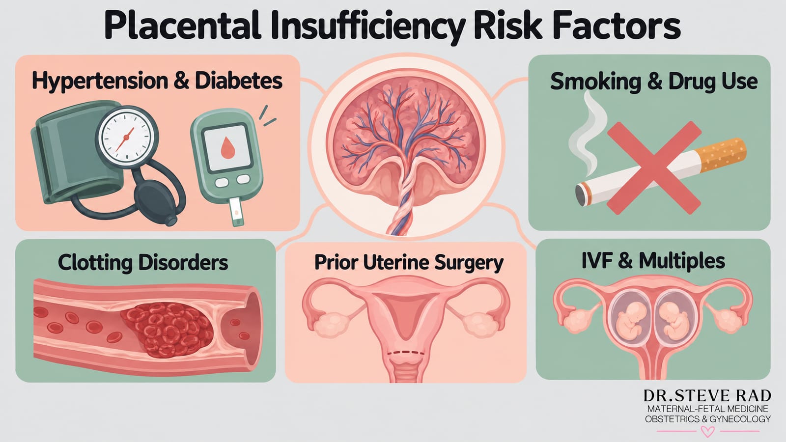
Pre-eclampsia or chronic hypertension
Diabetes
Smoking or illicit drug use
Blood clotting disorders
Prior uterine surgery or C-section, which can affect placental implantation and function
Pregnancies conceived through IVF, which carry a modestly elevated risk of placental complications
Carrying multiples, where placental demand is significantly higher
Almost 4% of pregnancy complications include a placental condition.
If you have been told your baby is measuring small, your fluid levels are low, or your Doppler results are abnormal, please do not navigate that news alone. A high-risk OB specialist can put a clear, personalized monitoring plan in place – and that plan can make all the difference for you and your baby.
Placenta accreta spectrum: accreta, increta, and percreta
Placenta accreta spectrum (PAS) disorders occur when the placenta attaches abnormally deeply into the uterine wall. They are among the most serious rare placenta problems, and their incidence is rising directly alongside C-section rates and IVF use, because uterine scarring creates the conditions for abnormal implantation.
Placenta accreta – the placenta attaches too deeply to the uterine wall (most common form, roughly 1 in 500 pregnancies)
Placenta increta – the placenta embeds into the uterine muscle wall
Placenta percreta – the placenta grows through the uterus and potentially into nearby organs such as the bladder
PAS disorders are typically diagnosed by ultrasound, with placental lakes or loss of the normal clear zone between placenta and uterine wall as key findings. MRI may be used to assess invasion depth. The standard management for significant accreta spectrum is a planned Cesarean hysterectomy, performed by a multidisciplinary team prepared for major hemorrhage and blood transfusion. In carefully selected cases where future fertility is a priority, conservative management may be considered, but this carries substantial risk and requires close specialist oversight.

The major consequences of these three conditions are that the placenta cannot come out after the baby is delivered and attempts to remove it result in heavy bleeding.
Using ultrasound, the perinatologist can make an accurate diagnosis before delivery. With a high-risk OB, these complications and risks can typically be managed.
Retained placenta
Separate from the accreta spectrum, a retained placenta occurs when the placenta fails to deliver naturally after birth. This is life-threatening without prompt treatment. There are three removal methods:
Manual removal by the physician, which carries an infection risk
Medication that relaxes the uterus to allow expulsion
Breastfeeding, which stimulates uterine contractions that can expel the placenta
Vasa previa
Vasa previa is one of the rarest placenta problems, occurring in approximately 1 in 2,500 births. It happens when fetal blood vessels from the placenta or umbilical cord run unprotected across the entrance to the birth canal. If those vessels rupture during labor, fetal hemorrhage can be rapid and fatal.
Vasa previa is often asymptomatic in the first trimester but may present with sudden heavy second-trimester bleeding. Because there are no warning signs in many cases, proactive screening is essential – especially for patients with a history of low-lying placenta, IVF conception, or a succenturiate (accessory) lobe placenta, all of which raise the risk. The gold-standard screening tool is transvaginal ultrasound with color Doppler, which can visualize the vessels crossing the cervix. This is typically performed between 18 and 20 weeks as part of a detailed anatomy scan, and again in the third trimester to confirm the diagnosis and guide delivery planning. A blood test can also rapidly confirm fetal blood in any vaginal bleeding, enabling timely intervention.
When a high-risk OB diagnoses it, outcomes improve significantly. Once your care team confirms vasa previa, they will discuss a structured management plan tailored to your situation. Recommended management includes hospitalization from around 30 to 34 weeks – allowing close monitoring of both mother and baby in a setting where an emergency response is immediately available – and planned Cesarean delivery between 34 and 37 weeks, before labor begins and before the risk of vessel rupture becomes critical. Delivering within this window is critical: studies show that when vasa previa is diagnosed prenatally and managed with planned Cesarean delivery, survival rates approach 97%, compared to outcomes that are far grimmer when the condition is discovered only at the time of rupture. This approach keeps fetal mortality very low. This is exactly the kind of high-stakes, detail-oriented care that a perinatologist like Dr. Rad is trained to provide – combining state-of-the-art ultrasound technology with the personal touch and 24/7 availability that anxious families deserve.
Circumvallate placenta
Circumvallate placenta occurs in about 1 to 2% of pregnancies, when the placenta folds back on itself during development. This reduces blood and nutrient flow to the baby, leading to fetal growth restriction, decreased amniotic fluid, and an elevated risk of placental abruption, miscarriage, or preterm birth. Ultrasound can diagnose or confirm it, sometimes with placental pathology review after delivery.
No direct treatment exists, but a structured monitoring protocol can minimize complications. Patients need additional growth scans and ultrasounds throughout pregnancy to track fetal development and amniotic fluid levels. If growth falls below acceptable thresholds, early delivery or emergency C-section may be necessary to protect the baby.
When to See a High-Risk OB for Placenta Problems
We understand that a placenta diagnosis can feel frightening, and knowing when to escalate your care is one of the most important things you can do for yourself and your baby. You should seek evaluation from a maternal-fetal medicine specialist if you experience any of the following:
Any vaginal bleeding during pregnancy, especially second trimester bleeding
A diagnosis of placenta previa, low-lying placenta, or vasa previa on ultrasound
Suspected placenta accreta spectrum, particularly with a history of C-section or uterine surgery
Reduced fetal movement or a uterus measuring small for gestational age
A diagnosis of placental insufficiency or intrauterine growth restriction
Sudden severe abdominal pain, which may signal placental abruption
Pregnancy conceived through IVF, which carries elevated PAS risk
Dr. Rad and his team at the Los Angeles Fetal and Maternal Care Center specialize in exactly these situations. We treat all patients as VIPs, ensuring you feel cared for and supported from your first consultation through delivery. We understand pregnancy can be a vulnerable and anxious time, and having the right specialist in your corner changes outcomes. The team is also available for second opinions and offers virtual consultations worldwide.
Call us at (844) 473-6100 or schedule your consultation online.
Call (844) 473-6100 or click here to schedule online
Placental conditions FAQs
What causes placental insufficiency?
Placental insufficiency, a condition where the placenta doesn’t function properly, can be caused by various factors. These include conditions such as high blood pressure, diabetes, autoimmune disorders, blood clotting disorders, and certain infections. Lifestyle factors such as smoking, drug use, and poor nutrition can also contribute to placental insufficiency. Additionally, maternal age, multiple pregnancies (such as twins or triplets), and certain genetic factors may increase the risk. Placental insufficiency can result in inadequate oxygen and nutrients reaching the fetus, leading to growth restriction, preterm birth, or other complications.
What causes placental abruption?
Placental abruption occurs when the placenta partially or completely separates from the uterine wall before delivery, which can lead to significant bleeding and other potential complications. The exact cause of placental abruption is not always clear, but it is often associated with high blood pressure, trauma to the abdomen, rapid uterine decompression, or certain risk factors like smoking, drug use, advanced maternal age, or previous incidences of placental abruption. These factors can disrupt the normal blood flow to the placenta, leading to its detachment from the uterine wall.
What is posterior placenta?
The posterior placenta refers to the placement of the placenta on the back wall of the uterus, towards the mother’s spine. Placental location can vary among pregnancies and can have implications for fetal development and maternal health. When the placenta is posterior, it typically doesn’t pose any significant risks on its own, but its position can affect the sensations a mother may feel during fetal movement. Additionally, the position of the placenta may influence the ease of visualization during ultrasound scans.
What is anterior placenta?
An anterior placenta refers to the placement of the placenta on the front wall of the uterus, closest to the mother’s abdomen. Like the posterior placenta, the location of the placenta can vary among pregnancies. Having an anterior placenta may impact a woman’s experience of feeling fetal movements.
Additionally, during ultrasound examinations, the anterior placenta may sometimes partially obstruct the view of the fetus, requiring the sonographer to adjust the scanning technique to obtain clear images.
What is battledore placenta?
A battledore placenta, also known as marginal cord insertion, is a rare variation in which the umbilical cord attaches to the edge or margin of the placenta instead of the center. This condition is typically considered benign and doesn’t usually cause significant problems during pregnancy. However, in some cases, a battledore placenta may be associated with an increased risk of complications such as restricted fetal growth or placental abruption, particularly if the cord insertion is very close to the placental edge.
Can circumvallate placenta go away?
Circumvallate placenta is a rare condition where the fetal membranes fold back upon themselves near the edge of the placenta, creating a raised ring-like structure. This condition is typically diagnosed during pregnancy through ultrasound examinations. Unlike other placental variations, circumvallate placenta does not resolve during pregnancy. It remains present throughout the duration of the pregnancy. While circumvallate placenta itself may not cause significant problems, it has been associated with an increased risk of complications such as placental abruption, preterm birth, and fetal growth restriction.
What causes low lying placenta?
A low-lying placenta, also known as placenta previa, occurs when the placenta implants low in the uterus, near or covering the cervix.
While most cases of low-lying placenta detected early in pregnancy may resolve on their own as the uterus grows and expands, some may persist and require close monitoring throughout pregnancy due to the risk of vaginal bleeding, preterm birth, or other complications.