Most parents assume 4D ultrasound is simply a “better” 3D ultrasound – but for high-risk pregnancies, the difference between these technologies can mean catching a fetal heart defect weeks earlier than a standard scan would. That distinction is not a marketing detail. It is a clinical one, and it matters enormously depending on your situation.
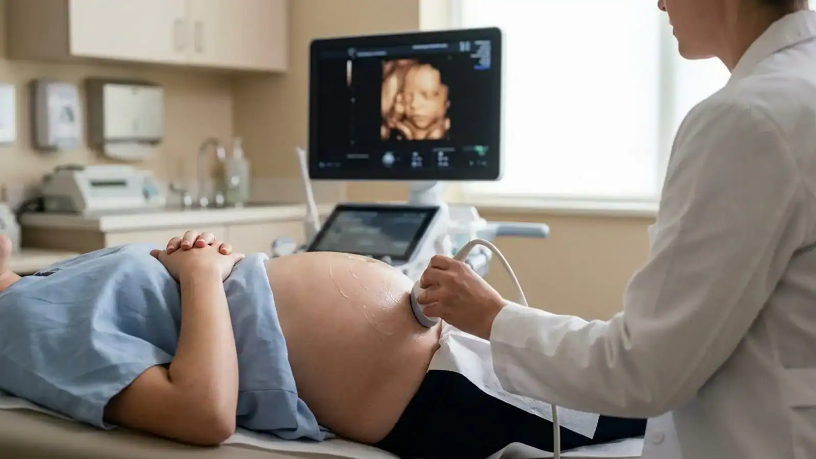
We understand pregnancy can be a vulnerable and anxious time. If you are trying to decide which type of ultrasound is right for you, or if your provider has recommended advanced imaging, you deserve a clear explanation from someone who uses this technology diagnostically every day – not just for keepsake photos. Dr. Steve Rad is a board-certified maternal-fetal medicine specialist voted Los Angeles’ best Maternal/Fetal specialist six years in a row by LA Magazine peers — and this guide reflects what he sees and acts on in clinical practice.
What follows is a maternal-fetal medicine perspective on how 2D, 3D, and 4D ultrasound actually work — why the distinction matters far more for high-risk patients than for keepsake shoppers, and what specific questions to ask before booking any advanced scan in Los Angeles or anywhere in Southern California.
2D, 3D, and 4D Ultrasound: What Each Technology Actually Does
All ultrasound technology works on the same fundamental principle: a transducer emits high-frequency sound waves that bounce off internal structures and return as echoes, which a computer converts into an image. What differs between 2D, 3D, and 4D is how those sound waves are collected and processed. The clinical implications of that difference are significant — and they are not always explained clearly to patients navigating a high-risk pregnancy. Dr. Steve Rad, a board-certified maternal-fetal medicine specialist voted Los Angeles’ best Maternal/Fetal specialist six years in a row by LA Magazine peers, uses all three modalities diagnostically every day — and here’s his clinical perspective on what each one actually does, and when each one matters most.
2D ultrasound sends sound waves in a single flat plane, producing the familiar black-and-white cross-sectional image. It shows internal structures — bones, organs, fluid — in a slice format. This remains the clinical workhorse of obstetric care because it penetrates tissue deeply and provides clear views of the anatomy that matters most for diagnosis.
3D ultrasound uses multiple angled sound wave sweeps captured simultaneously and reconstructed by software into a three-dimensional still image. Instead of a flat slice, you see the surface contours of the fetus — the curve of a cheek, the shape of a nose, the position of fingers. The image is static, like a photograph.
4D ultrasound adds time to that 3D reconstruction. The system captures and renders those three-dimensional images in rapid sequence — typically 20 to 30 frames per second — producing a live, moving video of the fetus. You can watch your baby yawn, stretch, or open and close their eyes in real time. For a maternal-fetal medicine specialist, that real-time motion is not just emotionally powerful. It is diagnostically essential.
If you are already in a high-risk pregnancy and wondering whether your current imaging is sufficient, schedule a consultation with Dr. Rad — we are happy to review your scan history and advise on next steps.
The One Key Difference Between 3D and 4D Ultrasound (It’s Not Just About Image Quality)
Parents researching 3D vs 4D ultrasound — what is the difference, exactly? — almost always find answers that focus on image quality or price. From a maternal-fetal medicine perspective, that framing misses the point entirely. The clinically relevant distinction is motion, and understanding it changes how you evaluate any advanced imaging recommendation during pregnancy.
What is the difference between 3D and 4D ultrasound?
The single most important difference is motion. A 3D ultrasound produces a detailed still image of the fetal surface at one moment in time. A 4D ultrasound produces a continuous, real-time video of fetal movement and behavior. Both use the same three-dimensional rendering process — the “4th dimension” is simply time added to the equation. So when patients ask about 3D vs 4D ultrasound and what the difference means for their care, the answer is: it depends entirely on what your specialist needs to observe.
For parents, this means 4D scans feel more like meeting your baby. For clinicians, it means something more specific: the ability to observe fetal cardiac motion, limb movement patterns, facial muscle activity, and swallowing behavior as they happen. A structural anomaly that might be missed in a single static frame can become visible when you watch the fetus move through multiple positions over several minutes.
Is a 4D ultrasound better than a 3D ultrasound?
Not categorically. Each has specific clinical applications where it performs best. 3D ultrasound excels at detailed surface rendering of facial structures, neural tube assessment, and skeletal evaluation — areas where a high-resolution still image gives the specialist time to analyze fine detail. Published research in Three- and Four-Dimensional Ultrasound in Obstetric Practice confirms that 3D ultrasound provides additional diagnostic value for facial anomalies, neural tube defects, and skeletal malformations because of its surface-rendering capability.
4D ultrasound is superior when the clinical question involves movement: cardiac function, fetal behavioral assessment, or real-time guidance during procedures. The right tool depends entirely on what your specialist needs. At Dr. Rad’s practice, we use both modalities together, selecting the appropriate mode based on gestational age and the specific clinical concern.
What Is HD Live Ultrasound – and Why Does the Machine Matter?
Beyond standard 3D and 4D, a newer generation of technology has changed what is diagnostically possible. HD Live ultrasound – available on GE ultrasound systems – applies a simulated light source to the three-dimensional rendering, creating images with depth, shadow, and texture that closely resemble a photograph of a newborn. The result is not just more emotionally striking. It reveals surface detail that standard 3D rendering can obscure.
5D ultrasound takes this further by incorporating automated measurement and analysis tools directly into the rendering software, reducing operator dependency and improving reproducibility of key fetal measurements. These are not consumer upgrades. They are clinical tools that require specialist training to interpret correctly.
What sets Dr. Rad’s practice apart is not the equipment alone — it is the combination of state-of-the-art GE HD Live technology and the interpretive expertise of a board-certified maternal-fetal medicine specialist. Ultrasound image quality is never just about the machine. A cutting-edge GE system operated by a trained perinatologist produces fundamentally different diagnostic information than the same technology used in a keepsake studio setting, where no physician is present to interpret what is on screen. To see what our advanced 3D and 4D ultrasound exams look like in practice, our ultrasound page walks through each modality in detail.
When Is Each Type of Ultrasound Used During Pregnancy?
When is the best time to get a 3D or 4D ultrasound during pregnancy?
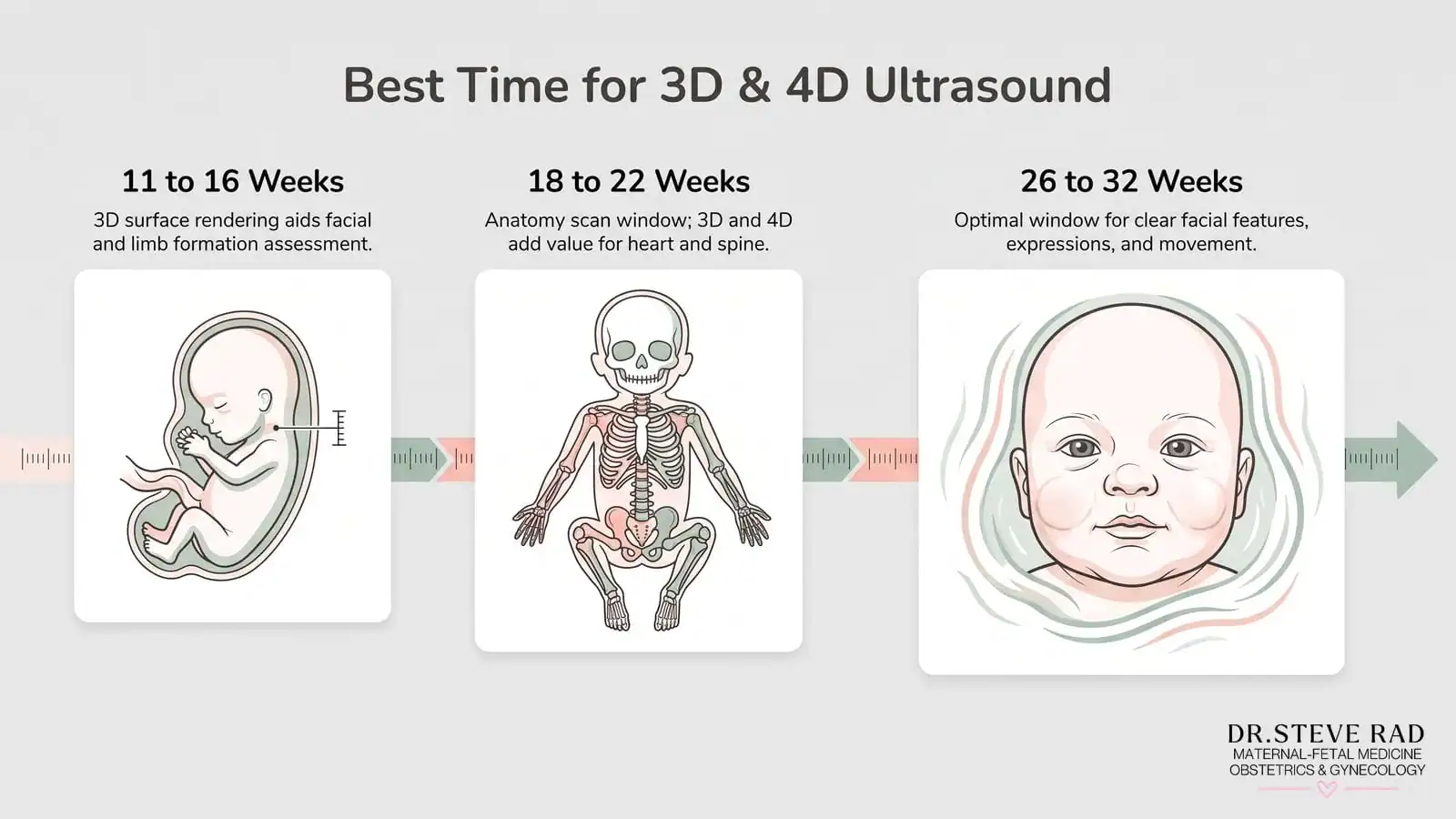
Gestational age significantly affects image quality and clinical utility for each modality. Here is how we approach timing in our practice:
11 to 16 weeks: This is the window for early fetal scans, where we can assess nuchal translucency, early cardiac structure, and gross anatomical development. At this stage, 2D remains the primary tool, but 3D surface rendering can add valuable information about facial development and limb formation. Early fetal diagnosis at this gestational age is a key capability that distinguishes a maternal-fetal medicine specialist from a general OB/GYN.
18 to 22 weeks: The anatomy scan window. 2D ultrasound is the standard, but 3D and 4D imaging add significant value for evaluating facial structures, the spine, and cardiac anatomy. A fetal echocardiogram – a detailed assessment of the fetal heart using specialized ultrasound protocols – is typically performed in this window for patients with elevated cardiac risk.
26 to 32 weeks: The optimal window for 3D and 4D surface imaging. The fetus has enough subcutaneous fat to produce clear surface detail, and there is still sufficient amniotic fluid surrounding the face for an unobstructed view. This is when the images parents associate with 3D/4D scans – detailed facial features, expressions, movement – are most reliably obtained.
If you are unsure which imaging window applies to your situation — or whether your current scan schedule is sufficient for a high-risk pregnancy — schedule a consultation with Dr. Rad to review your options.
Why 4D Ultrasound Is a Clinical Tool, Not Just a Keepsake, for High-Risk Pregnancies
Consider a patient referred to us at 20 weeks with a suspected ventricular septal defect identified on a routine anatomy scan at her general OB’s office. Using 4D ultrasound with color Doppler flow mapping, we can observe blood flow through the fetal heart in real time, assess the defect’s size and location, and evaluate whether it affects cardiac function. That real-time picture lets us stratify risk immediately—distinguishing a small, hemodynamically insignificant defect that may close on its own from one that requires a neonatal surgical plan in place before delivery. A still image simply cannot provide that information.

For patients managing high-risk pregnancy conditions – including gestational diabetes, pre-eclampsia, fetal growth restriction, or chromosomal abnormalities – 4D ultrasound gives us behavioral and functional data that changes clinical management. We can assess fetal breathing movements, gross body movements, and tone as part of a biophysical profile. We can monitor placental blood flow in real time. These are not bonding experiences. They are diagnostic assessments that directly inform care decisions.
One patient came to us after two previous pregnancy losses, both attributed to undetected cardiac anomalies at standard anatomy scans. Using our GE HD Live 4D system at 14 weeks — earlier than most practices attempt detailed cardiac assessment — we identified an early structural concern with the fetal outflow tract. That finding triggered an immediate referral to a pediatric cardiologist, who joined the care team months before delivery to map out a surgical plan. We coordinated the birth at a facility equipped for immediate neonatal cardiac intervention, and the family arrived at delivery with a team already in place rather than facing an emergency no one had prepared for. When detection happens early enough to change the plan, it changes everything.
If you are managing a high-risk pregnancy and are unsure whether your current imaging is giving your care team the full picture, schedule a consultation to discuss whether advanced 4D imaging is right for your situation.
Can a 4D Ultrasound Detect Birth Defects? What MFM Specialists Look For
Can a 4D ultrasound detect birth defects or fetal abnormalities?
Yes, with important qualifications. Advanced 4D ultrasound in the hands of a trained maternal-fetal medicine specialist can identify a wide range of fetal anomalies, including cardiac defects, cleft lip and palate, neural tube defects, skeletal dysplasias, and abdominal wall abnormalities. The published literature confirms that 3D ultrasound specifically adds diagnostic value for facial anomalies and skeletal malformations beyond what 2D imaging alone provides.
However, no ultrasound modality – 2D, 3D, or 4D – detects every possible fetal anomaly. Detection rates depend on gestational age, fetal position, maternal body habitus, amniotic fluid volume, and, critically, the operator’s expertise and equipment quality. A 4D scan at a keepsake studio is not diagnostic. It is an imaging session performed without clinical context, without a specialist interpreting the findings, and without the protocols that govern a medical ultrasound examination.
Keepsake Studio vs. Maternal-Fetal Medicine Specialist: Understanding the Difference
What is the difference between a keepsake ultrasound and a medical ultrasound?
A keepsake ultrasound is performed primarily for the emotional experience of seeing your baby in three dimensions. The operator is typically not a licensed medical professional, the session is not governed by clinical protocols, and findings are not interpreted or documented as part of your medical record. Organizations including the American Institute of Ultrasound in Medicine (AIUM) have consistently cautioned against non-medical ultrasound sessions because they involve fetal exposure to ultrasound energy without clinical justification.
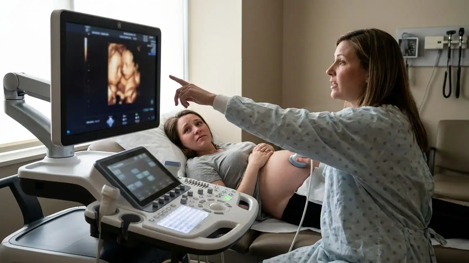
At Dr. Rad’s practice, every advanced ultrasound is performed within a clinical framework. Findings are documented, interpreted, and communicated to the patient with the full context of their obstetric history and risk profile.
A medical ultrasound performed by a maternal-fetal medicine specialist is diagnostic. It follows established protocols, is performed by or under the direct supervision of a board-certified physician, and produces findings that are integrated into your care plan. The images may look similar on screen. The clinical value is not comparable.
We treat all patients as VIPs, ensuring you feel cared for and supported — but that concierge experience is built on a foundation of clinical rigor, not just comfort. For patients who want the emotional experience of seeing their baby in HD alongside the reassurance of a specialist’s eyes on every frame, our practice offers both in the same appointment. And for high-risk patients who need more monitoring but find frequent clinic visits difficult, we also offer at-home prenatal care—including ultrasound imaging—so specialist-level oversight is never out of reach.
How much does a 3D or 4D ultrasound cost in Los Angeles?
Pricing varies significantly depending on where you go and what you receive. Keepsake studios in Los Angeles typically charge between $50 and $200 for a session, reflecting the fact that no physician is present, no clinical interpretation is provided, and the images are not integrated into your medical record.
A diagnostic 3D or 4D ultrasound performed by or under the supervision of a board-certified maternal-fetal medicine specialist is a medical procedure and is priced accordingly. For patients with a documented high-risk condition, insurance often covers advanced ultrasound in part by insurance, particularly when ordered as part of a biophysical profile, fetal echocardiogram, or growth assessment. We recommend contacting your insurance provider directly to understand your coverage before booking.
At Dr. Rad’s practice, we are transparent about costs from the outset and will help you understand what your insurance may cover. If you have questions about pricing or coverage for advanced imaging, schedule a consultation and our team will walk you through your options.
What to Ask Before Booking a 3D or 4D Ultrasound in Los Angeles
Not all advanced ultrasound services in Los Angeles are equivalent—and the differences aren’t always obvious from a clinic’s website or pricing page. In our experience, patients who ask the right questions before booking are far better positioned to get imaging that actually informs their care. Here is what we recommend asking, and why each question matters.
Who performs and interprets the scan? A board-certified OB/GYN or maternal-fetal medicine specialist should perform the scan or directly supervise it. Sonographers alone, without physician oversight, cannot provide a clinical interpretation. This is the single most important question you can ask — because the same image means something entirely different depending on who is reading it.
What equipment is being used? GE HD Live technology represents the current standard for diagnostic 3D/4D imaging. Ask specifically about the machine model and whether HD Live rendering is available. While GE HD Live systems are commercially available, the diagnostic value they deliver depends entirely on the expertise of the specialist interpreting the images — not the machine alone. The difference between a GE HD Live scan read by a board-certified perinatologist and the same scan performed at a keepsake studio without physician oversight is not equipment. It is a difference in clinical meaning.
Is the scan integrated into your prenatal care record? A standalone keepsake session produces images. A clinical ultrasound produces a report that becomes part of your medical record and informs your ongoing care. For high-risk patients especially, that documentation is not a formality — it is the thread that connects every specialist involved in your pregnancy.
Can early fetal scans be performed? If you have a high-risk pregnancy, the ability to perform diagnostic imaging as early as 11 to 16 weeks is clinically significant. Not all providers offer this capability. Dr. Steve Rad, voted Los Angeles’ best Maternal/Fetal specialist six years in a row, routinely performs early fetal scans as part of comprehensive high-risk pregnancy monitoring — because in perinatology, earlier answers almost always lead to better outcomes.
Frequently Asked Questions about 3D and 4D ultrasounds
Are 3D and 4D ultrasounds safe during pregnancy?
When qualified medical professionals perform them for clinically indicated reasons, 3D and 4D ultrasounds use the same sound-wave technology as standard 2D scans and carry the same safety profile. The AIUM and major obstetric organizations consider diagnostic ultrasound safe when used appropriately. The concern raised about keepsake studios is not that ultrasound itself is dangerous, but that prolonged, non-clinically indicated exposure without medical oversight introduces unnecessary risk without diagnostic benefit. In a clinical setting with a trained specialist, the exposure is controlled, purposeful, and proportionate to the information being sought.
Do I need a 3D or 4D ultrasound if I have a high-risk pregnancy?
For many high-risk pregnancies, advanced 3D and 4D imaging is not just beneficial – it is a meaningful upgrade over standard 2D scanning for specific diagnostic questions. If your pregnancy involves a suspected fetal cardiac anomaly, a structural concern identified on a routine scan, a history of chromosomal abnormalities, or conditions like fetal growth restriction, the real-time functional data from 4D ultrasound and the surface detail from 3D imaging can directly change clinical management. Your maternal-fetal medicine specialist will determine which modalities are appropriate based on your specific risk profile and gestational age. Explore the full range of high-risk pregnancy conditions we manage to understand where advanced imaging fits into your care.
What is an HD or 5D ultrasound and how does it differ from 4D?
HD Live ultrasound applies a simulated directional light source to a standard 3D/4D rendering, producing images with realistic shadow and depth that reveal surface detail more clearly than conventional 3D rendering. 5D ultrasound incorporates automated measurement tools and AI-assisted analysis into the rendering process, reducing operator variability and improving the reproducibility of key fetal measurements.
Both represent advances beyond standard 4D imaging, but they require specific equipment – such as the GE HD Live system – and specialist training to use diagnostically. They are not widely available, which is why the equipment a provider uses matters when you are evaluating where to have advanced prenatal imaging. For patients in Los Angeles seeking this level of technology, our at-home prenatal ultrasound care brings this capability directly to you.
How much does a 3D or 4D ultrasound cost in Los Angeles?
Pricing varies significantly depending on where you go and what you receive. Keepsake studios in Los Angeles typically charge $100 to $250 for a session, but that price reflects an imaging experience—not a diagnostic examination. There is no physician interpretation, no clinical report, and no integration with your prenatal care record.
A 3D or 4D ultrasound performed at a maternal-fetal medicine practice like Dr. Rad’s is a different service entirely. Pricing depends on gestational age, scan complexity, and whether it is part of a broader clinical assessment such as a fetal echocardiogram or biophysical profile. Many advanced ultrasound examinations are covered in full or in part by insurance when there is a documented clinical indication — which is almost always the case for patients managing high-risk pregnancies.
We encourage patients to schedule a consultation to discuss their specific situation. Our team will clarify what is clinically indicated, what your insurance is likely to cover, and what out-of-pocket costs to expect — so there are no surprises. That transparency is part of the concierge, VIP experience we provide to every patient.