When expecting a child, parents and healthcare providers alike make your baby’s health a top priority. Monitoring your baby’s heart for early abnormalities is crucial, and a fetal echocardiogram plays a vital role. By examining your baby’s heart while they are still in the womb, we can diagnose fetal cardiac problems before your baby is even born, giving you more time to work with experts and develop a care plan tailored to your baby’s needs.
We understand pregnancy can be a vulnerable and anxious time. Our goal is to make sure you feel informed, supported, and never alone in this process. That is the Dr. Rad difference.
What is a fetal echocardiogram?
A fetal echocardiogram (also called a fetal echo) is a specialized prenatal ultrasound that provides a comprehensive view of your unborn baby’s heart. While a traditional fetal ultrasound shows your baby’s overall development, a fetal echo focuses exclusively on the fetal heart. A trained cardiac sonographer collects detailed data on your baby’s blood flow, blood vessels, heart chambers, heart valves, and fetal heart rate, including heart rhythm.
The scan provides crucial insights into your baby’s cardiovascular health. Because we detect irregularities early in pregnancy, you have plenty of time to plan treatments or interventions. Working with pediatric cardiology experts, Dr. Rad can prepare a full postnatal care plan for your baby before they even arrive. Some congenital heart defects require immediate intervention at birth, and knowing about these conditions ahead of time means the right specialists are ready.
Who needs a fetal echocardiogram?
The heart is the body’s most important muscle. Congenital heart disease (CHD) can put your baby’s long-term health at risk, so most parents want to know in advance if their baby may have a heart condition. Not every pregnancy requires a fetal echo, but if you have risk factors for CHD, Dr. Rad may recommend a complete evaluation of the fetal heart.
A fetal echocardiogram may be recommended if:
You have a family history of congenital heart defects or congenital heart disease.
A previous child was born with a structural heart defect or CHD.
Another prenatal test revealed a chromosomal or genetic disorder, or a birth defect was suspected on a routine anatomy scan.
You have certain maternal medical conditions such as pre-gestational diabetes, lupus, rubella, or phenylketonuria (PKU).
You conceived through in vitro fertilization (IVF) or other assisted reproductive technologies.
You were exposed to teratogenic medications during pregnancy, including certain anti-seizure drugs, lithium, or retinoids.
Your baby showed an irregular heart rhythm or abnormal heart rate on a routine ultrasound.
There is an abnormal amount of amniotic fluid (polyhydramnios or oligohydramnios).
You have a multiple pregnancy (twins or more) with shared circulation concerns.
If you are unsure whether you need a fetal echo, Dr. Rad will review your full history and guide you. Many patients are referred by their OB-GYN, but you can also request a consultation directly.
When is a fetal echocardiogram done?
A fetal echocardiogram is most commonly performed between 18 and 22 weeks of pregnancy. This window is considered the sweet spot because the baby’s heart structures are developed enough to be clearly visualized, yet there’s still plenty of time to plan next steps if anything unexpected is found. Think of it as getting the most complete picture at exactly the right moment.
That said, timing isn’t always one-size-fits-all. If a concern is identified earlier – for example, during a first-trimester screening or an advanced fetal ultrasound – Dr. Rad may recommend an earlier evaluation, sometimes as soon as 14 to 16 weeks. While the images at this stage can be more limited, a preliminary look can still provide valuable reassurance or prompt closer monitoring.
On the other end of the spectrum, a fetal echocardiogram can also be performed later in the third trimester if a new concern arises or if a follow-up scan is needed to track a previously identified finding. For patients managing high-risk pregnancies, serial echocardiograms – meaning more than one scan over the course of the pregnancy – may be recommended to monitor how the baby’s heart is developing over time.
Dr. Rad will always discuss the ideal timing for your situation during your consultation, ensuring every scan is scheduled when it will give you and your care team the most useful, actionable information. That personal touch is at the heart of everything we do – that is the Dr. Rad difference.
What to expect during your fetal echocardiogram appointment
Knowing exactly what will happen during your appointment can ease a great deal of anxiety. Here is a step-by-step walkthrough of what a typical fetal echo visit looks like at Dr. Rad’s practice.
Arrival and preparation
Unlike a standard obstetric ultrasound, you do not need to arrive with a full bladder for a fetal echocardiogram. You can eat and drink normally beforehand. When you arrive, a member of our team will welcome you, review your medical history, and answer any questions before the scan begins.
Positioning and gel application
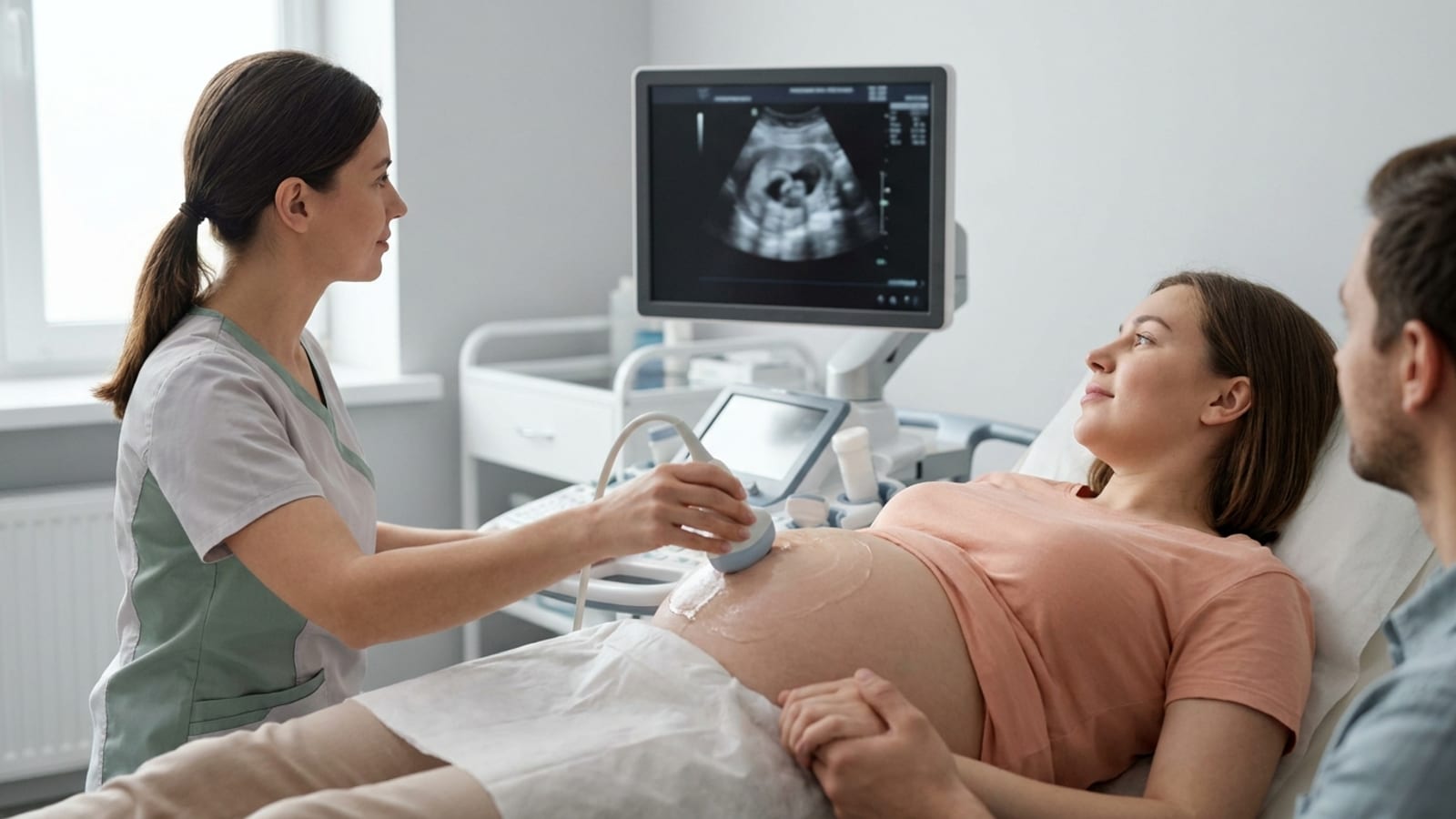
You will lie comfortably on an exam table. The cardiac sonographer will apply a warm, water-based gel to your abdomen. This gel helps the ultrasound transducer make good contact with your skin and ensures clear sound wave transmission. The transabdominal approach, in which the transducer is moved gently across your abdomen, is the standard method for second-trimester fetal echocardiograms. In earlier gestational ages or when image quality requires it, a transvaginal approach may be used, which is also painless and safe.
The scan itself
The cardiac sonographer will systematically image your baby’s heart from multiple angles. Key views include the four-chamber view, which shows all four chambers of the heart simultaneously, as well as outflow tract views and Doppler assessments of blood flow through the valves and great vessels. The sonographer is measuring the size of the heart chambers, the thickness of the walls, the direction and speed of blood flow, and the rhythm of the heartbeat. The scan typically takes 45 to 60 minutes, though it may take a little longer if your baby is in a position that makes imaging more challenging.

The tech will apply gel to your abdomen and move the ultrasound transducer across your stomach to capture the necessary images. In addition to images of your baby’s heart, we also use color Doppler to capture the direction of blood flow.
The test usually takes about 30 minutes to an hour but may take up to two hours if your baby is in a difficult position.
The images and videos will be reviewed by a pediatric cardiologist to evaluate any heart abnormalities.
After the scan
Once the sonographer completes the imaging, Dr. Rad reviews the results and, when appropriate, consults with a pediatric cardiologist. You will receive a thorough explanation of the findings, what they mean for your baby, and the recommended next steps. We never send you home with a report and no context. Our team will walk you through every finding, answer every question, and make sure you leave feeling informed and supported.
Your care team: who performs and reviews the scan
Who will perform my fetal echocardiogram?
One of the most reassuring things you can know before your appointment is exactly who will be in the room and what role each person plays. A fetal echocardiogram isn’t a standard ultrasound – it’s a specialized study that brings together a skilled team working on your behalf.
Cardiac Sonographer
With Dr. Rad, your fetal echocardiogram is performed by an experienced cardiac sonographer – a technician with advanced training specifically in fetal heart imaging. They operate the state-of-the-art ultrasound equipment, carefully capturing detailed views of your baby’s heart structures, valves, and blood flow patterns. This takes patience and precision, so the scan typically takes 45-60 minutes.
Dr. Rad, perinatologist and maternal-fetal medicine specialist
Once the images are captured, Dr. Steve Rad – a renowned maternal-fetal medicine specialist and award-winning perinatologist – reviews the findings in full. His deep expertise in high-risk pregnancies means he can interpret even subtle findings with confidence and explain them to you in plain, reassuring language.
Pediatric cardiologist
If the results raise concerns, Dr. Rad coordinates directly with pediatric cardiologists and other specialists to build a clear care plan before your baby arrives. You’re never left to navigate those conversations alone. That collaborative, concierge approach – where your care team communicates seamlessly so you don’t have to chase answers – is what sets this experience apart from a generic ultrasound appointment.
From the sonographer capturing the images to Dr. Rad guiding your next steps, everyone involved focuses on one thing: giving you and your baby the best possible outcome. That is the Dr. Rad difference.
What will the fetal echocardiogram results show?
After your fetal echocardiogram, the results will generally fall into one of two categories: normal or abnormal. Either way, knowing what the scan found gives you and your care team the information needed to move forward with confidence.
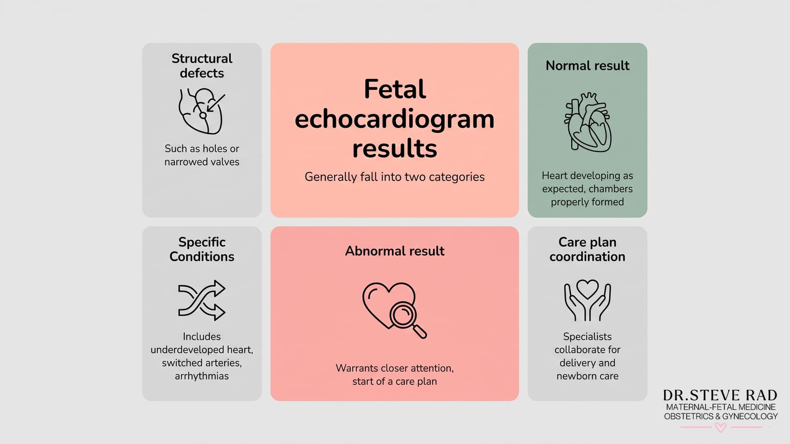
A normal result means the baby’s heart appears to be developing as expected – the four chambers are properly formed, the valves are opening and closing correctly, and blood is flowing in the right direction. For most families, this is exactly the reassurance they were hoping for.
An abnormal result means the scan has detected something that warrants closer attention. This might include conditions such as:
Structural defects – such as holes between the heart’s chambers (septal defects), narrowed or malformed valves, or vessels that are connected incorrectly
Hypoplastic left or right heart syndrome – where one side of the heart is underdeveloped
Tetralogy of Fallot – a combination of four heart abnormalities that affect blood flow
Transposition of the great arteries – where the two main arteries leaving the heart are switched
Arrhythmias – irregular heart rhythms that may need monitoring or treatment
It’s important to understand that an abnormal result is not a crisis – it’s the beginning of a care plan. As a renowned maternal-fetal medicine specialist, Dr. Rad works closely with pediatric cardiology experts to ensure that if a heart condition is detected, your family is fully prepared well before your baby arrives. That means coordinating the right specialists, discussing delivery planning, and making sure your newborn receives the care they need from day one.
Some findings may be minor and resolve on their own, while others require surgical intervention after birth. Whatever the result, Dr. Rad will walk you through every detail in plain language – because that personal touch and 24/7 availability are the Dr. Rad difference.

Call (844) 473-6100 or click here to schedule online
What happens after a fetal echocardiogram?
If your fetal echo results are normal, no further cardiac imaging is typically needed, though Dr. Rad will continue to monitor your pregnancy as part of your overall care plan.
If the results show an abnormality, the next steps depend on the nature and severity of the finding:
We may schedule a repeat fetal echocardiogram to monitor a finding over time or obtain clearer images as the baby grows.
Referral to a pediatric cardiologist for a specialist consultation and detailed counseling about the specific condition detected.
Coordination with a perinatologist or maternal-fetal medicine (MFM) specialist to manage any associated pregnancy risks.
Genetic counseling or additional testing, such as amniocentesis, if a chromosomal condition is suspected alongside the cardiac finding.
Delivery planning at a hospital with a neonatal intensive care unit (NICU) and pediatric cardiac surgery capabilities, so the right team is in place the moment your baby arrives.
Dr. Rad coordinates all of these referrals and conversations on your behalf, so you are never left navigating the system alone.
Are fetal echocardiograms safe?
Yes, fetal echocardiograms are completely safe for both mother and baby. The procedure uses the same sound-wave technology as a standard obstetric ultrasound, so there is no radiation exposure. The sound waves are non-invasive and painless, and clinicians have used them safely for decades. Fetal echocardiography has no known risks or side effects, and major medical organizations, including the American Heart Association, support its use as a diagnostic tool when clinically indicated.
The procedure does not require sedation, does not involve any needles or incisions, and causes no discomfort beyond the mild sensation of the gel and transducer on your abdomen. It is one of the safest diagnostic tools available in prenatal medicine.
We understand that when your provider recommends additional testing during pregnancy, it can feel unsettling. That is why we want to be clear: a fetal echocardiogram is not a high-stakes intervention – it is simply a more detailed look at your baby’s heart using the same gentle, non-invasive ultrasound technology you have already experienced. The sound waves used during the scan pass harmlessly through tissue, creating real-time images without radiation or chemical exposure. Because this technology has been refined and studied over several decades, its safety profile is exceptionally well established across millions of pregnancies worldwide.
From a practical standpoint, there is nothing to prepare for in terms of physical risk. You will lie comfortably on an exam table, warm gel will be applied to your abdomen, and Dr. Rad or a trained sonographer will gently move the transducer across your skin. The scan itself typically takes between 45 and 60 minutes – longer than a routine ultrasound – simply because the heart is a complex structure and a thorough evaluation takes time. That extra time shows diligence, not cause for concern. Many patients find the experience reassuring once they can see their baby’s heart beating clearly on the screen.
Have concerns about your pregnancy? Contact Dr. Rad
Dr. Rad is one of Los Angeles’s premier high-risk obstetricians and maternal-fetal medicine specialists, and he understands the nuances of fetal echocardiogram results with a depth that comes from years of experience managing complex pregnancies. He knows how important early, accurate diagnoses are, and how profoundly they can improve health outcomes for babies and peace of mind for families.
For parents facing high-risk pregnancies or who have concerns about their baby’s heart health, the guidance and expertise of a specialized perinatologist are invaluable. Dr. Rad and his world-class Maternal-Fetal Medicine team at Los Angeles Fetal and Maternal Care Center are dedicated to providing comprehensive care and support throughout every stage of your pregnancy.
Their expertise in high-risk obstetric issues, combined with a compassionate, personalized approach, ensures you receive the best possible care. Whether you have questions about fetal echocardiography, congenital heart disease, high-risk pregnancy management, or simply need support during this crucial time, Dr. Rad and his team are here to help.
Call us at (844) 473-6100 or schedule your consultation online. We are currently accepting new patients.
We are conveniently located for patients throughout Southern California and the Los Angeles area at locations in or near Beverly Hills, Santa Monica, West Los Angeles, West Hollywood, Culver City, Hollywood, Venice, Marina del Rey, Malibu, Manhattan Beach, Newport Beach, Irvine, and Downtown Los Angeles. We also offer in-home prenatal care and a fly-in program for out-of-town and international patients. Dr. Rad even travels to patients who need him throughout the U.S. and around the world.
If you cannot make it to Dr. Rad in person, he also offers virtual consultations worldwide.
Call (844) 473-6100 or click here to schedule online
Frequently asked questions about fetal echocardiograms
Is a fetal echocardiogram the same as a regular pregnancy ultrasound?
No. A routine obstetric ultrasound gives your care team a broad overview of your baby’s development — size, position, organ formation, and amniotic fluid levels. A fetal echocardiogram is a dedicated, in-depth study of the heart alone. It uses the same safe sound-wave technology, but a specialized cardiac sonographer performs the scan; it takes longer (45–60 minutes), and it captures detailed views of the heart’s chambers, valves, and blood flow that a standard ultrasound simply cannot provide.
Is a fetal echocardiogram safe for my baby?
Yes — completely. A fetal echocardiogram uses sound waves, not radiation, to image your baby’s heart. The technology is non-invasive and painless, and clinicians have used it safely for decades. The procedure itself carries no known risks to you or your baby. If you have any concerns, Dr. Rad and our team are always happy to walk you through the science before your appointment.
How accurate is a fetal echocardiogram?
A fetal echocardiogram is the most accurate prenatal tool available for evaluating the fetal heart. In experienced hands, it can detect most significant structural heart defects before birth. That said, no test is 100% perfect — image quality can be affected by factors such as the baby’s position, gestational age, and maternal body habitus. If the initial images are inconclusive, Dr. Rad may recommend a follow-up scan at a later gestational age for a clearer picture.
Will my insurance cover a fetal echocardiogram?
Coverage varies by insurance plan and the clinical indication for the scan. When a fetal echocardiogram is ordered because of a documented risk factor — such as a family history of congenital heart disease, a chromosomal finding, or a maternal medical condition — it is typically covered as a medically necessary procedure. Our team will work with you to verify your benefits and navigate the prior authorization process before your appointment, so you won’t be surprised.
What if the fetal echocardiogram finds a problem?
Finding an abnormality on a fetal echocardiogram is understandably frightening, but it is important to remember that early detection is a powerful advantage — not a verdict. Many congenital heart defects are manageable, and knowing about them before birth gives your care team time to plan. Dr. Rad will coordinate directly with pediatric cardiologists and, where needed, arrange delivery at a hospital equipped with a neonatal intensive care unit (NICU) and pediatric cardiac surgery capabilities. You will never be left to navigate those conversations alone — that is the Dr. Rad difference.
Can a fetal echocardiogram miss a heart defect?
While a fetal echocardiogram is highly sensitive, some defects — particularly small ventricular septal defects or mild valve abnormalities — may not be visible until later in pregnancy or after birth. This is why Dr. Rad may recommend serial echocardiograms (more than one scan over the course of your pregnancy) for high-risk cases, and why postnatal cardiac evaluation remains important even after a normal fetal echo result.